
Table of Contents
What Is a Talar Dome Fracture?
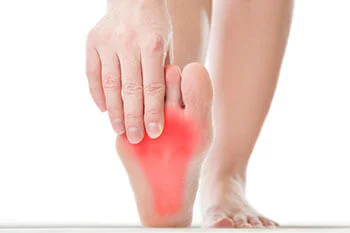
Signs & Symptoms
The signs and symptoms of a talar dome lesion may include:
- Lasting pain deep in the ankle that is worse with activity
- Clicking or catching feeling in the ankle
- Ankle locking or a feeling that the ankle is “giving out.”
- Ankle swelling
Diagnosis
Nonsurgical Treatment
- Immobilization. Sometimes a period of immobilization with a cast or cast boot is used to allow the lesion to heal. An ankle brace may needed if the ankle is unstable to prevent recurrent ankle sprains and further injury to the ankle joint.
- Oral medications: Nonsteroidal anti-inflammatory drugs (NSAIDs), such as ibuprofen, can be helpful to reduce pain and inflammation.
- Steroid Injections: Steroid injections into the joint using a form of cortisone can be performed to also reduce pain and inflammation.
- Physical therapy: Once the lesion has had a chance to heal, physical therapy can also be helpful to reduce pain and swelling.
Surgical Treatment
If conservative treatment does not resolve pain, surgical treatment may be recommended. The procedure is dependent on the size of the lesion, but in most cases, ankle arthroscopy is recommended.
Arthroscopy of the ankle is a less invasive surgical technique performed through small incisions that give full access to a joint. This involves placing a telescopic camera (the diameter of a drinking straw) and small tools into the joint to clean up the lesion and stimulate healing while watching the surgery on a monitor. The obvious advantages to arthroscopy over traditional techniques are the smaller incisions that permit early range of motion (often in the same day), as well as a reduction in postoperative recovery time with less pain and swelling.
Typically, patients are non-weightbearing for 1-2 weeks to permit the small incisions to fully heal, slowly increasing activities as tolerated for the next 4-6weeks, until a return to full activities can be accomplished.
After surgery, in most instances, we recommend early range of motion activities, especially if scar tissue was found within the ankle. This early motion helps to prevent return of the adhesions and promote faster recovery.
If the ankle arthroscopy fails to relieve pain or the talar dome lesion is very large, a type of Cartilage Resurfacing Surgery may be recommended.